Research Article - Annals of Biological Research ( 2018) Volume 9, Issue 1
Triphynel methane dyes are recalcitrant owing to their xenobiotic nature and exhibit high resistance to degradation processes. In the present study, two different bacteria Gram positive “Bacillus cereus” and Gram negative” Pseudomonas earoginosa” isolated from soil samples contaminated with industrial effluent, collected from Jeddah industrial city, Saudi Arabia, were analyzed for Triphynel methane dyes, Malachite Green (MG), and Crystal Violet (CV) biodegradation. The physicochemical parameters such as aeration, incubation periods, adding carbon and nitrogen sources, pH, and temperature affecting the biodegradation of (MG) and (CV) were tested. The maximum (MG) and (CV) degradation was found to be the maximum at 95.2%, 96.3% by B. cereus and P. aeruginosa at shaking conditions after 3 days of incubation. The optimum temperature was 37°C for decolorizing by both bacterial strains. The optimum pH to decolorize (MG) and (CV) was 7 by B. cereus and it was 8 to decolorize (MG) and (CV) by P. earoginosa. Studying the effect of dye concentration showed that B. cereus recorded the highest percentage at (100 mg/L) with 91.2% and 88.3% for MG and CV respectively, and P. aeruginosa were able to decolorize malachite green and crystal violet at a the same concentration up to 90%. Glucose was the best carbon source for malachite green and crystal violet which increased the rate of decolorization up to 90% after 72 hours of incubation by B. cereus, followed by fructose. P. aeruginosa recoded a high percentage for MG and CV at 95.3% and 91% respectively. Peptone was the best nitrogen source for the decolorzation of malachite green while yeast extract was the best source of nitrogen which showed high percentage in decolorization of crystal violet. UV-Visible spectrum analysis demonstrated disappearing of major peaks of (MG) and (CV) at 590 nm and 619 nm, at visible area and formation a new peaks at UV area
Physicochemical parameters, Malachite green, Crystal violet, Decolorization
Triphenylmethane dyes are xenobiotic and aromatic compounds which used widely in many industrial purposes such as biological stains, textile paper printing, and dye manufacturing industries [1].
The triphenylmethane dye, crystal violet (Tris(4-(dimethylamino) phenyl)methylium chloride), has been extensively used in laboratories as a biological stain and in various commercial textile processes as a dye [2]. Malachite green (N-methylated diaminotriphenylmethane) is an organic compound that has been used extensively for dyeing silk, wool, jute, leather, ceramic and cotton [3]. It is highly soluble in water and has also been used in aquaculture industry as a fungicide, parasiticide and disinfectant [4].
Crystal violet and Malachite green has been classified as a recalcitrant molecule, they resist to biodegrade by some microorganisms [5]. Therefore, most of biological treatment for industrial wastewater under aerobic and an aerobic conditions are not sufficient to remove all degradative by products. These dyes are potentially toxic, mutagenic and carcinogenic [6].
In contrast, many reports have been studied the decolorization of triphenylmethane dyes by several microorganisms such as fungi, algae, and bacteria, actinomycetes, and yeast.
Parshetti [7] investigated the biodegradation of Malachite Green by Kocuria rosea MTCC 1532 which capable to produce malachite green reductase and DCIP reductase. Chiing-Chang Chen [8] studied the biodegradation of crystal violet by Pseudomonas putida which was effective to degrade CV up to 80%. Olukanni [9] investigated the biodegradation of Malachite Green by extracellular Laccase producing Bacillus thuringiensis RUN1.
Also, Ali et al. [10] studied the Decolorization and degradation of malachite green by A. flavus and Alternaria solani, and they found Both the species were able to decolorize different concentrations of malachite green (10 to 50 M) almost completely (>96%) within 6 days. Jadhav and Govindwar [11] investigated the biotransformation of malachite green by Saccharomyces cerevisiae MTCC 463.
The aim of this study is to focus on decolorization of two different dyes related to TPM class by using aerobic bacteria Pseudomonas aeruginosa and Bacillus cereus.
Chemicals
Crystal violet (C25H30CIN3) (Figure 1) and malachite green (C23H25N2) 2.3C2H2O4 (Dye content MIN.90%) Figure 2 were purchased from Loba Chemia India.
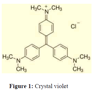
Figure 1: Crystal violet.
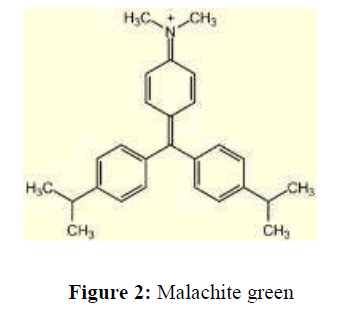
Figure 2: Malachite green.
Medium
Mineral salts basal medium is used for assaying degradation activities of bacterial strains, the composition of this media is as follow (g/L): NaH2PO4 (0.235), MgSO4.7H2O (0.07), CaCl2 (0.014), FeCl3. 6H2O, (0.001) glucose was used as carbon source (10) and Yeast extract was used as nitrogen source (5.0), the medium was autoclaved at 121°C and 1atm for 40 min. In the following, the Malachite green and crystal violet with final concentration of 100 mg added to the Erlenmeyer flask containing MSM medium separately. Eventually, the results were analyzed after 48 hours by UV-Vis spectroscopy.
Bacteria
Two different bacterial species “Bacillus cereus” and “Pseudomonas aeruginosa” known as aerobic bacteria were obatianed from Alborg Medical Laborites, Jeddah, Saudi Arabia for the present study maintained in nutrient agar slants until further investigation. The pure culture of “B. cereus” and “P. aeruginosa” were grown on nutrient agar at 37°C for 24 h.
Decolorization assay
All decolorization experiments were performed in three stages: At first, after autoclavation of the nutrient broth medium, a loopful of bacteria grown on nutrient agar was inoculated in 250 mL Erlenmeyer flask containing 100 mL nutrient broth and incubated at 37°C for 24 h. Secondly, after 24 h of incubation, bacterial inoculum of B. cereus and P. aeruginosa was transferred to the MSM contained concentration of 50 mg/L for each dye. After three days of incubation the bacteria with tested dyes. Aliquot was centrifuged at 1000 rpm for 10 minutes by refrigerated centrifuge to separate the bacterial cell mass, clear supernatant which was used to measure the decolorization of MG and CV and determine by measuring the absorbance at 590 nm and 619 nm using a UV-1800 UV/VIS Spectrophotometer (RAYLEIGH, Beijing Beifen-Ruili Analytical Instrument (Group) Co., Ltd.) Abiotic control (without bacteria inoculation) was used for measuring. Finally, the percentage decolorization was calculated using the following equation:
Where, Initial absorbance: Initial dye conc. (mg/L), Final absorbance: Residual dye conc. (mg/L).
Effect of shaking and static conditions during different incubation periods on the decolorization efficiency of bacteria
The cultures containing Malachite green and Crystal violet were incubated in a shaking incubator and in static conditions at different periods 24, 48, 72 hours, three replicates from each treatment were used.
Effect of dye concentration on the decolorization efficiency of bacteria
The concentrations of malachite green and crystal violet added into the MSM were at 100 mg/L, 200 mg/L, 500 mg/L, 1000 mg/L, 1500 mg/L and 2000 mg/L. For each concentration of the mixture, three replicates from each treatment were used.
Effect of temperature on the decolorization efficiency of bacteria
The cultures containing Malachite green and Crystal violet were incubated at different temperatures which were 25°C, 30°C 37°C, 40°C, 45°C, and 50°C respectively. For every bacterial culture incubated under different temperatures, three replicates from each treatment were used.
Effect of pH value on the decolorization efficiency of bacteria
The pH of the MSM was adjusted accordingly using 0.1 N sodium hydroxide or 0.1 N hydrochloric acid to pH 5, 6, 7, 8, 9 and 10. For each broth of differing pH, three replicates from each treatment were used.
Effect of different carbon sources on the decolorization of bacteria
Four different carbon sources such as glucose, fructose, Sucrose, and Starch were added separately to 250 ml elementary flask containing 100 ml of the MSM and incubated at 37°C for 48 hours. Three replicates from each treatment were used.
Effect of different nitrogen sources on the decolorization of bacteria
Four different nitrogen sources such as yeast extract, casein, peptone, and Sodium Nitrate were added separately to 250 ml elementary flask containing 100 ml of the MSM and incubated at 37°C for 48 hours. Three replicates from each treatment were used.
Data were calculated as mean ± standard deviation (SD).
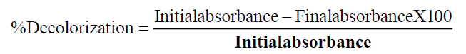
Measurement of biodegradation of Crystal Violet and Malachite green by using UV-spectrophotometer analysis
Figure 3: Spectroscopic analysis of dyes before decolorization of Crystal violet.
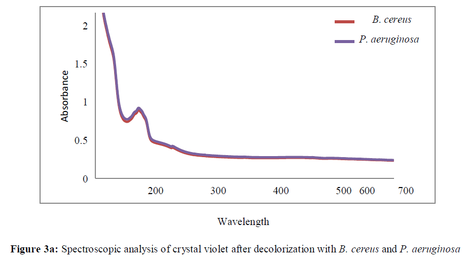
Figure 3a: Spectroscopic analysis of crystal violet after decolorization with B. cereus and P. aeruginosa.
The maximum absorbance peaks of crystal violet and malachite green were at 590 nm and 618 nm respectively as shown in Figures 3 and 4, After treatment by B. cereus and P. aeruginosa, the maximum absorbance peaks for both dyes decreased significantly. A new peaks then appeared at ultra violet area for crystal violet treated with B. cereus and P. aeruginosa (Figures 3a and 4a).
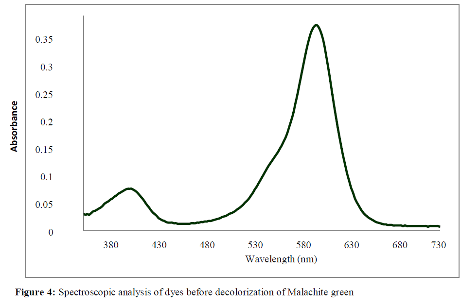
Figure 4: Spectroscopic analysis of dyes before decolorization of Malachite green.
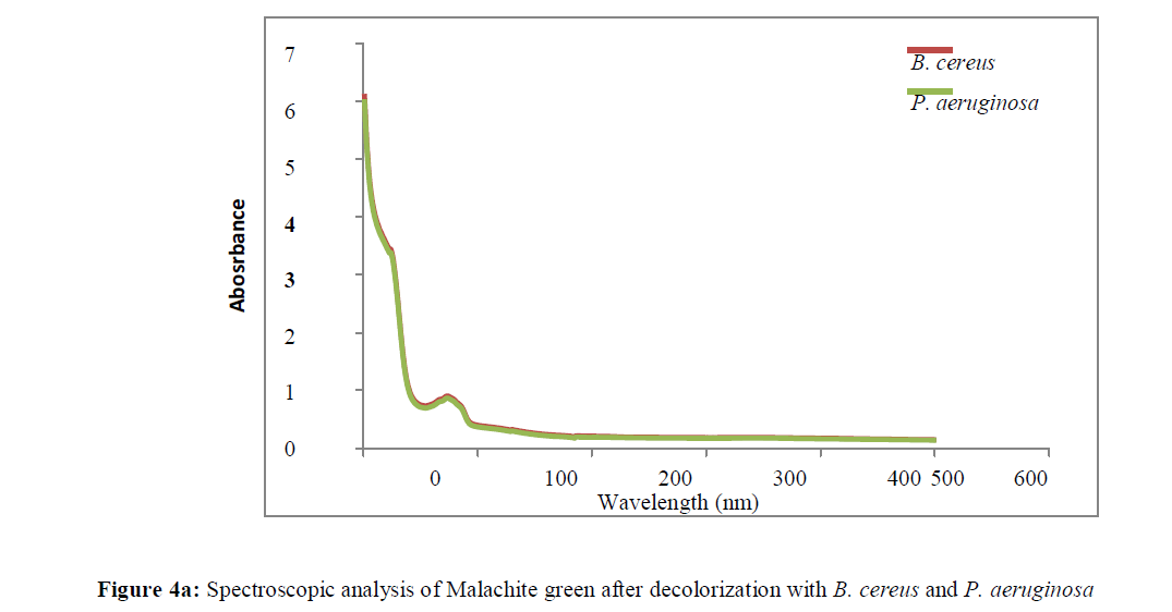
Figure 4a: Spectroscopic analysis of Malachite green after decolorization with B. cereus and P. aeruginosa.
These peaks most likely corresponded to the formation of colorless derivatives upon degradation of crystal violet and malachite green as reported by Jang, et al. (2005) and Ye, et al. (2007).
The study was conducted to examine the effects of aeration on the level of efficiency of dyes decolorization. Two different conditions, which were the shaking and static conditions, were set. Based on the results shown in Table 1a malachite green was better decolorized by B. cereus under the shaking condition in all the three periods of incubation.
| Bacteria | Malachite Green | ||||||
|---|---|---|---|---|---|---|---|
| Static Conditions | Shaking Condition | ||||||
| Incubation periods | |||||||
| 24 | 48 | 72 | 24 | 48 | 72 | ||
| Bacillus cereus | 38.3 ± 1.3 | 86.5 ± 2.6 | 85.7 ± 1.6 | 2.64 ± 0.6 | 87.2 ± 0.9 | 92.4 ± 0.9 | |
| Pseudomonas aeruginosa | 39.3 ± 0.7 | 83.8 ± 1.6 | 86.3 ± 1.3 | 41.3 ± 1.3 | 88.4 ± 0.9 | 95.7 ± 0.4 | |
Table 1a: Effect of shaking and static conditions on decolorization of Malachite green at different incubation periods.
The level of decolorization of malachite green under the shaking condition was the highest at 92.4% while under the static condition; it was only 85.7% after 72 hours of incubation. Whereas P. aeruginosa showed a good activity to decolorize MG in shaking conditions better than static conditions. The highest percentage of decolorization was 95.7% in shaking conditions comparing with the percentage of static conditions which recorded 86.3% after 72 hours of incubation.
Based on Table 1b, the decolorization pattern of crystal violet was similar to that of malachite green under the shaking and static conditions. The levels of decolorization of crystal violet were much higher under the shaking condition compared to the static one in all the three periods of incubation. The level of decolorization of crystal violet reached its peak for B. cereus and P. aeruginosa at 95.2% and 96.3% respectively after 72 hours of incubation under the shaking condition. The percentage of static conditions was less than shaking conditions. B. cereus and P. aeruginosa recorded 86.2% and 85.4% respectively after 72 hours of incubation period.
| Bacteria | Crystal Violet | |||||
|---|---|---|---|---|---|---|
| Static Conditions | Shaking Condition | |||||
| Incubation periods | ||||||
| 24 | 48 | 72 | 24 | 48 | 72 | |
| Bacillus cereus | 37.4 ± 1.5 | 77.3 ± 0.9 | 86.2 ± 1.1 | 43.1 ± 1 | 89 ± 2.8 | 95.2 ± 1 |
| Pseudomonas aeruginosa | 44.1 ± 1 | 81 ± 1.3 | 85.4 ± 0.8 | 48.3 ± 0.5 | 91.2 ± 1.7 | 96.3 ± 1.1 |
Table 1b: Effect of shaking and static conditions on decolorization of Crystal violet at different incubation periods.
Generally P. aeruginosa known as an aerobic bacteria which depends on dissolved oxygen for its growth [12,13]. Under the shaking condition, the aeration in the flask was promoted, hence increasing the amount of dissolved oxygen in it. Besides, the nutrients were also distributed evenly in the mixture under the shaking condition which in turn maximized the nutrient uptake by bacteria [14]. Hence, the bacterial growth was increased which in turn contributed to a high level of decolorization of the dyes. Likewise, a moderate activity of efficiency in the decolorization of dyes under the static condition was due to a low level of dissolved oxygen in the mixture. Moreover, the bacterial cells might settle to the bottom of the flasks under this condition and became oxygen depleted. This resulted in a slower rate of cell growth and in turn affected the dye decolorization process [15].
| Bacteria | Temperature ( °C) | |||||||||||
|---|---|---|---|---|---|---|---|---|---|---|---|---|
| Malachite Green | Crystal violet | |||||||||||
| 25 | 30 | 37 | 40 | 45 | 50 | 25 | 30 | 37 | 40 | 45 | 50 | |
| Bacillus | 34 | 91.6 | 98.5 | 78 | 56.6 | 35.5 | 39.4 | 85.4 | 95.1 | 91.2 | 37 | 20.8 |
| cereus |  ± 2 |  ± 1.5 |  ± 0.9 |  ± 1.8 |  ± 1.2 |  ± 1.1 |  ± 0.75 |  ± 1.6 |  ± 1.6 |  ± 1.5 |  ± 1.4 |  ± 1.6 |
| Pseudomonas | 27.7 | 56.6 | 91.5 | 89.4 | 65 | 52.1 | 51.6 | 85.7 | 93.2 | 88.6 | 47 | 27.5 |
| aeruginosa |  ± 1.4 |  ± 0.9 |  ± 0.9 |  ± 1.4 |  ± 1.4 |  ± 1.8 |  ± 1.7 |  ± 0.9 |  ± 0.9 |  ± 1 |  ± 1.5 |  ± 0.9 |
Table 2: Effect of different temperature degrees on decolorization of MG and CV dyes.
Data (Table 2) showed that, Media inoculated by B. cereus showed the best decolorization at 37°C for MG and CV at 98.5% and 95.1%, while incubation at 37° C was found to be optimum and has the efficiency to decolorize MG and CV at 91.5% and 93.2% by P. aeruginosa. Percentages of decolorization at minimum temperature 25°C and maximum temperature at 50°C for both bacterial strains were lower compared with the optimum temperature for each bacteria at the same incubation period (72hours). Generally, temperature degree plays a vital role in the growth of bacteria especially at optimum temperature. Conversely, at a higher temperature of 37° C, the oxygen solubility decreases and eventually causes a reduction in the metabolic activity of the aerobic natured B. cereus and P. aeruginosa [16]. Since dye degradation is a metabolic process, shifting to higher temperature will lower the decolorization activity of bacteria.
| bacteria | PH | |||||||||||
|---|---|---|---|---|---|---|---|---|---|---|---|---|
| Malachite Green | Crystal violet | |||||||||||
| 5 | 6 | 7 | 8 | 9 | 10 | 5 | 6 | 7 | 8 | 9 | 10 | |
| Bacillus cereus | 16.2 | 33.4 | 94.4 | 74.6 | 68.5 | 57 | 19.5 | 22.5 | 96.3 | 86.2 | 74.6 | 50.7 |
|  ± 1.6 |  ± 1.7 |  ± 2.1 |  ± 1.1 |  ± 1.4 |  ± 1.4 |  ± 1.4 |  ± 0.8 |  ± 1.1 |  ± 0.4 |  ± 1.5 |  ± 1 | |
| Pseudomonas | 19.3 | 40.2 | 59.3 | 93.8 | 80.4 | 59.6 | 24.5 | 43.4 | 51.2 | 92.3 | 79.4 | 41 |
| aeruginosa |  ± 1.2 |  ± 1.3 |  ± 0.9 |  ± 0.8 |  ± 1.2 |  ± 0.8 |  ± 1 |  ± 1.1 |  ± 1.1 |  ± 1 |  ± 0.8 |  ± 1.2 |
Table 3: Effect of different pH on decolorization of MG and CV dyes.
Based on Table 3, at the alkaline pH, B. cereus also results showed the highest percentage of decolorization rate at 94.4% and 96.3% for MG and CV respectively at pH 7 after 72 hours of incubation period. This result agrees with Santhi and Nalina [17] which reported that B. cereus recorded the best decolorization at pH 7. While P. aeruginosa recorded the highest percentage of decolorization at 93.8% and 92.3% after 72 hours of incubation for MG and CV respectively. pH plays a vital role in the ionization and binding of substrates, solubility of the nutrients required by the bacteria for their growth and decolorization ability, it is necessary to be optimized. An optimum pH for malachite green was varied and depends on to the bacterial strain used by different investigators. Earlier, Wu et al. [18] reported similar observations for decolourization of a triphenylmethane dye by Pseudomonas otitidis. Ji’ai [19] reported that the color of MG remove at pH 6 and temperature 38 by A. xylosoxidans. Generally, Pseudomonas sp. required optimum pH of 8.0 [20] for malachite green whereas, Sphingomonas sp. required pH 9.0 [21].
The effects of pH values on the levels of decolorizing activities of P. aeruginosa and B. cereus on crystal violet and malachite green were investigated with pH values ranging from 5 to 10. P. aeruginosa and B. cereus exhibited low levels of decolorization activities on crystal violet and malachite green at pH 5, pH 6, pH 9 and pH 10. Bacteria at lower pH-value (5) degree can’t grow well, therefore; the decolorization efficiency was affected by low pH and recorded low results. Bacteria at pH (9-10) shows moderate activity to decolorize the color of MG and CV. triphenylmethane dyes are more toxic at a lower pH and this has made the bacterial cells more susceptible to their toxic effects [22].
The effect of dye concentrations on the decolorization activities of B. cereus and P. aeruginosa was investigated with different concentrations of crystal violet and malachite green. Table 4 shows the dye removal efficiency of crystal violet and malachite green with the initial dye concentration ranging from 100 mg/L to 1500 mg/L by B. cereus and P. aeruginosa After 72 hours of incubation.
| Bacteria | Dye Concentration( mg/L ) | |||||||||
|---|---|---|---|---|---|---|---|---|---|---|
| Malachite Green | Crystal violet | |||||||||
| 100 | 200 | 500 | 1000 | 1500 | 100 | 200 | 500 | 1000 | 1500 | |
| Bacillus cereus | 91.2 ± 1.7 | 88.6 ± 2.9 | 70.7 ± 1.5 | 33.1 ± 1.3 | 25.5 ± 1.8 | 88.3 ± 1.8 | 84.1 ± 1.7 | 61.3 ± 0.97 | 30.3 ± 1.1 | 22.3 ± 1.6 |
| Pseudomonas aeruginosa | 95.8 ± 1.5 | 91.4 ± 1.6 | 73.2 ± 1.5 | 36.8 ± 1.6 | 24.3 ± 1.5 | 93.3 ± 1.7 | 88.7 ± 1.5 | 68.1 ± 1.7 | 34.5 ± 1 | 19 ± 1.6 |
Table 4: Effect of different Concentrations of MG and CV dyes (mg/L) on decolorization rates.
All results showed that increasing of decolorization rate based on initial dyes concentrations, dyes with low concentration (mg/L) biodegrade faster than dyes with high concentrations.
The percentage of dye decolorization for MG and CV decreased with the increasing of dye concentration, B. cereus recorded the highest percentage at (100 mg/L) with 91.2% and 88.3% for MG and CV respectively.
P. aeruginosa was able to decolorize malachite green and crystal violet at a concentration of 100 mg/L up to 90%. Reduced dye decolorization activities were observed at the dye concentrations of 500 mg/L to 1500 mg/L.
According to Fischer, et al. [23] the increasing of concentration of crystal violet and malachite green lead to the decreasing of the decolorization efficiency by bacteria due to the toxicity of the triphenylmethane dyes6 Triphenylmethane dyes of high concentration exhibited bacteriostatic effect which makes the lag phase period longer by inhibiting cellular metabolic activities and cell growth of bacteria. On the other hand, Malachite green which has only two dimethyl groups shows lower antibacterial activity compared to crystal violet which has three dimethyl groups [24].
Various carbon sources such as glucose, fructose, sucrose and starch were added separately to MSM at concentration (10 g/L), as shown in Table 5 among these carbon sources, glucose was the best carbon source for malachite green and crystal violet which increased the rate of decolorization up to 90% after 72 hours of incubation by B. cereus, followed by fructose. P. aeruginosa recoded a high percentage for MG and CV at 95.3% and 91% respectively. This result agrees with Ouranusi and Mbah [25].
| Bacteria | Carbon Sources | |||||||
|---|---|---|---|---|---|---|---|---|
| Malachite Green | Crystal violet | |||||||
| Glucose | Fructose | Sucrose | Starch | Glucose | Fructose | Sucrose | Starch | |
| Bacillus cereus | 93.6 | 90.6 | 86.5 | 81.9 | 94.4 | 78.7 | 87.4 | 76.8 |
|  ± 0.8 |  ± 1 |  ± 1.1 |  ± 1.5 |  ± 0.7 |  ± 1.7 |  ± 1.1 |  ± 1.05 | |
| Pseudomonas | 95.3 | 75.4 | 82.5 | 89.4 | 91 | 86.4 | 82.3 | 80.3 |
| aeruginosa |  ± 1 |  ± 1 |  ± 2.1 |  ± 1.8 |  ± 1.5 |  ± 0.7 |  ± 1.5 |  ± 1.1 |
Table 5: Effect of different carbon sources on decolorization rates of MG and CV dyes.
| Bacteria | Nitrogen Sources | |||||||
|---|---|---|---|---|---|---|---|---|
| Malachite Green | Crystal violet | |||||||
| Yeast extract | Casein | Peptone | Ammonium Nitrate | Yeast extract |
Casein | Peptone | Ammonium Nitrate | |
| Bacillus cereus | 90.2 | 71.6 | 96.5 | 70.2 | 96.6 | 60.8 | 91 | 85.2 |
|  ± 2.09 |  ± 0.8 |  ± 0.9 |  ± 1.1 |  ± 0.9 |  ± 1.3 |  ± 0.9 |  ± 0.9 | |
| Pseudomonas | 89.6 | 82.6 | 1.69 | 66.6 | 92.1 | 76.7 | 88.3 | 77.2 |
| aeruginosa |  ± 2.25 |  ± 1 |  ± 1.1 |  ± 1.08 |  ± 1.4 |  ± 1.4 |  ± 1 |  ± 1.4 |
Table 6: Effect of different Nitrogen sources on decolorization rates of MG and CV dyes.
Various nitrogen sources such as yeast extract, casein, peptone and Ammonium nitrate were added separately to MSM at concentration (5 g/L), as show in Table 6. The results of adding these substances to the medium showed that peptone was the best nitrogen source for the decolorzation of malachite green. B. cereus and P.aeruginosa showed a good activity at 96.5% and 93.1% respectively. This result agrees with Santhi and Nalina [17] and Omar [26] which found peptone was the best nitrogen source to decolorize MG. Yeast extract was the best source of nitrogen which showed high percentage in decolorization of crystal violet. B. cereus and P. aeruginosa recorded 96.6% and 92.1% respectively after 72 hours of incubation. In a similar optimization studies carried out by Ji’ai [19], peptone and beef extract were found to be enhancing the malachite green decolourizing ability of Achromobacter xylosoxidans [27-31].