Research Article - Annals of Biological Research ( 2017) Volume 8, Issue 2
This study involving four public restaurants randomly selected in Ado-Ekiti metropolis was aimed at isolating, characterizing and comparing the susceptibility of bacteria associated with equipment’s used in food processing to conventional antibiotic discs and chloroform extract of Moringa oleifera. Swab samples were collected from equipment and environment used for food processing and were taken to the laboratory were microbiological analysis were carried out. Fifty nine bacteria isolates were obtained altogether. The bacteria isolated from these samples were Escherichia coli (15), Bacillus sp. (10), Serratia marcenscens (5), Staphylococcus aureus (11), Bacteriodes spp. (4), Citrobacter youngae (5), Klebsiella spp. (4), Enterobacter aerogenes (5). Multiple antibiotic resistances were discovered in some of the isolates when subjected to antibiotic susceptibility test using conventional antibiotic discs. Some phenotypic multiple antibiotic resistance pattern observed are AUG/CAZ/CRX/LIN/OXC/CXC and TET/AMX/COT/NIT/GEN/NAL/AUG among others. Majority of the isolates were susceptible to chloroform extract of M. oleifera with the exception of some isolates such as S. aureus, E. coli, Klebsiella sp. and Bacteroides sp. This study has shown the prevalence and resistance of microorganisms associated with public restaurants in Ado-Ekiti metropolis to conventional antimicrobial agents and the effectiveness of chloroform extract of M. oleifera in serving as an alternative antimicrobial agent. In conclusion, it is imperative for public restaurants to ensure that equipment, personnel’s and environment used/involved in food processing are very clean in order to reduce the occurrence of food borne microorganisms to the barest minimum
Swab samples, Antimicrobial agents, Multiple antibiotic resistance
According to FAO/WHO, food safety is the assurance that when food is consumed, it does not cause harm to human health and wellbeing [1]. Food safety is of utmost concern in the twenty-first century as a result of food borne illnesses which are widespread and continues to be a public health problem worldwide. Consequently, consumers are increasingly concerned about food safety and quality; and therefore demand more transparency in production and distribution of food [2,3].
Food borne illness of microbial origin is a major health problem associated with public foods [4,5]. Food borne diseases are common in developing countries because of the prevailing poor food handling and sanitation practices, inadequate food safety laws, weak regulatory systems, lack of financial resources to invest in safer equipment, and lack of education of food handlers [6]. According to Linda du and Irma [7], some factors contributing to outbreak of food borne diseases include unsafe sources of food items, contaminated raw food items, improper food storage, poor personal hygiene etc. Food service establishments and food handlers contribute to food borne illness outbreaks [8]. Approximately 10 to 20% of food-borne disease outbreaks are due to contamination by the food handler [9].
According to the Food Safety Certification Regulations in the United States, almost 75% of food borne illness outbreaks is assumed to be related to improper food handling practices by employees in food establishments. Some studies have shown that poor sanitary conditions of catering establishments favors the presence of pathogenic organisms like Campylobacter, Salmonella, Staphylococcus aureus, Bacillus cereus and Escherichia coli [10-13]. Most public eating places in Nigeria especially in the low cost areas are characterized by unsanitary conditions, including poor water supply and poor drainage systems, unsanitary waste disposal and overcrowding, resulting in poor personal and environmental hygiene [14]. Good personal hygiene and food handling practices are essential for preventing the transmission of pathogens from food handlers to the consumers [15].
The selection of the appropriate treatment to be used for food borne illnesses depends on the identification of the causal organism, and determining if specific therapy is available. Gastroenteritis requires fluid replacement and supportive care. Oral rehydration is required by patients who are mildly to moderately dehydrated, while intravenous therapy is used in severe cases of dehydration. The choice of therapy to be used is usually based on clinical signs and symptoms, organisms detected in the clinical specimens, the antimicrobial susceptibility tests and the appropriateness in treating with an antibiotic [16]. The treatment of food borne infection with antibiotics is however associated with the adverse effect of drug resistance by the microorganisms. According to CDC [17], the issue of drug resistance among microorganisms has become a global challenge with at least 2 million people acquiring serious infections with bacteria that are resistant to one or more of the antibiotics designed to treat those infections each year in the United States, resulting in about 23,000 death each year with many more deaths as a result of other conditions complicated by an antibiotic-resistant infection.
Moringa oleifera commonly referred to as Moringa is a highly nutritious evergreen or deciduous tree that usually grows up to 10 to 12 m in its height. M. oleifera has been used for various purposes [18,19]. Various parts of this plant such as the leaves, roots, seed, bark, fruit, flowers and immature pods act as cardiac and circulatory stimulants, possess anti-tumor, antipyretic, antiepileptic, anti-inflammatory, antiulcer, antispasmodic, diuretic, antihypertensive, cholesterol lowering, antioxidant, anti-diabetic, hepatoprotective, antibacterial and antifungal activities. Hence, they are being employed for the treatment of different ailments in the traditional system of medicine [19-21]. This study was therefore designed to investigate factors associated with food safety practices of food handlers working in food establishments in Ado-Ekiti metropolis, isolate and characterize major food contaminating microbes in public restaurants and carry out susceptibility tests using conventional antibiotic discs and extracts from Moringa oleifera.
Study area and population
This study was conducted using public restaurants in Ado-Ekiti, Ekiti State which is located between latitudes 7°34´ and 7°41´N of the equator and longitudes 5°11´ and 5° 6´E of the Greenwich meridian. The four public restaurants used in this study were randomly selected in Ado-Ekiti town.
Ethical consideration
Approval was sought and collected from the public restaurants before the commencement of the research.
Sampling technique
Samples were collected from various equipment’s such as tomato filter, grinding machines, tables used for cutting and other cooking equipment used for food processing in the different restaurants using swab sticks. All the samples taken were stored at 4°C using ice packs and were transported to the Microbiological Laboratory and were immediately analyzed.
Sample analysis
The swab sticks were inoculated into already sterilized and cooled nutrient broth immediately the samples arrived in the laboratory. The nutrient broth tubes were incubated aerobically at 37°C for 24 h after which sub culturing was done on different media such as blood agar, mannitol salt agar, EMB agar, MacConkey, Salmonella-Shigella agar and nutrient agar (all produced by Oxoid). Further sub culturing was carried out in order to purify the obtained bacterial isolates. The isolates were observed for presumptive identification based on their morphological characteristics, Gram staining and various biochemical tests that included catalase, oxidase, hydrogen sulfide production, motility, indole, methyl red, urea, Voges-Proskauer, growth on Triple sugar iron (TSI) agar and citrate utilization tests [22-26].
Plant sample collection
The Moringa leaves used in this research work were obtained from the Moringa plantation of the teaching and research farm of Afe Babalola University Ado-Ekiti (ABUAD), Nigeria.
Preparation of M. oleifera extracts
Fresh leaves of M. oleifera were obtained and air dried for weeks. After drying, the leaves were grinded to fine powder using an electric blender. One hundred gram of the powdered Moringa leaves was soaked into 500 ml of chloroform and left for 72 h at room temperature after which it was filtered using filter paper. M. oleifera extract was obtained by drying off the methanol using rotary evaporator.
Antibiotic susceptibility test
The isolates obtained from the samples were subjected to antibiotic susceptibility test following the Clinical and Laboratory Standards Institute (CSLI) method [27] using Kirby-Bauer disk diffusion test on Muller-Hinton agar (Oxoid CM0337 Basingstoke, England). Each isolate was inoculated into nutrient broth separately and incubated for 24 h at 37°C. The broth were streaked using sterile cotton swabs on Mueller-Hinton Agar plates. This was followed by aseptic placement of the antibiotic discs using sterile forceps. The plates were incubated aerobically at 37°C for 24 h, after which the zones of inhibition were measured and interpreted according to CSLI [27]. Antibiotics used for Gram negative isolates were Augmentin (30 μg), Ofloxacin (5 μg), Gentamicin (10 μg), Nalidixic acid (30 μg), Nitrofurantoin (200 μg), Amoxicillin (25 μg), Tetracycline (25 μg), while those used for Gram positive isolates were Augmentin (30 μg), Cotrimoxazole (25 μg), Cloxacillin (5 μg), Erythromycin (5 μg), Gentamicin (10 μg), Streptomycin (10 μg), Tetracycline (10 μg) and Chloramphenicol (10 μg).
Antimicrobial activity of Moringa extracts using agar-well diffusion method
Susceptibility of the isolated organisms to Moringa extracts was determined using agar well diffusion technique. Agar plate containing Muller-Hinton agar to which the suspension of the test bacteria was added was prepared using pour plate technique, 7 mm diameter wells were bored into the agar plates. The chloroform extracts were diluted, using DMSO as diluents concentrations (500-100 mg/ml) and were added to the wells. The plates were left at ambient temperature for 15 min and then incubated at 37°C for 24 h, after which the zones of inhibition were observed and recorded. A well containing DMSO was used as control.
Four public restaurants in Ado-Ekiti were used for this study. From these public food restaurants, twelve equipment and facilities used in food processing and the kitchen floor were swabbed for sample collection. The samples were obtained from tomato filter, stainless working table, meat cutting table, bean filter, grinding machine, cutlery used by consumers, toasting machine, pastry machine, rice section burner, baking table, chiller and the kitchen floor.
Fifty-nine bacteria isolates were obtained altogether with thirty eight being Gram negative while twenty one of the isolates were Gram positive. The bacteria isolated from these samples include Escherichia coli (15), Bacillus subtilis
(4), B. cereus (3), B. badius (1), B. coagulans (2), Serratia marcenscens (5), Staphylococcus aureus (11), Bacteriodes spp. (4), Citrobacter youngae (5), Klebsiella spp. (4), Enterobacter aerogenes (5).
The frequency of occurrence of the bacterial isolates obtained is shown in Figure 1, it was observed that E. coli had the highest frequency of occurrence among the isolates while B. badius had the least frequency of occurrence.
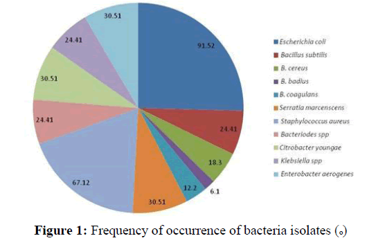
Figure 1: Frequency of occurrence of bacteria isolates (o)
The frequency of occurrence of isolates obtained per equipment is shown in Figure 2. Equipment’s such as grinding machine and tomato filter gave higher bacterial isolates (11 each) than other equipment’s such as baking table, toasting machine etc. used in food processing in the four public restaurants.
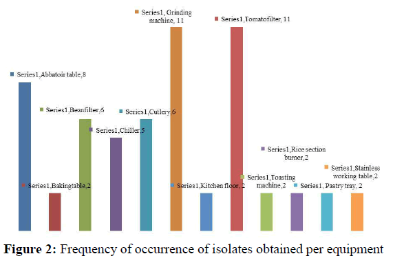
Figure 2: Frequency of occurrence of isolates obtained per equipment.
Figure 3 shows the number of each bacterial isolate obtained per equipment. It was observed that E. coli occurred more often in the chiller than any other equipment’s used for food processing.
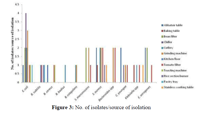
Figure 3: No. of isolates/source of isolation
The antimicrobial susceptibility test revealed that the bacterial isolates showed varying degree of susceptibility to the antibiotics used. Table 1 shows the susceptibility of the Gram positive isolates to the antibiotic discs used. It was observed that majority of the Gram positive isolates showed resistance to most of the antibiotics used. For instance, all the Gram positive isolates were resistant to augmentin, ceftazidime, cefuroxime, lincomycin, oxacillin and cloxacillin. Some of the Gram positive isolates were however susceptible to gentamicin (80.95%) and ofloxacin (100%). There were two major phenotypic multiple antibiotic resistance pattern observed among the Gram positive isolates. Seventeen of the isolates showed resistance to seven out of the eight antibiotic discs used with a phenotypic multiple antibiotic resistance pattern of AUG/CAZ/CRX/GEN/LIN/OXC/CXC, while four isolates were resistant to six out of the eight antibiotics tested against them with a phenotypic multiple antibiotic resistance pattern of AUG/ CAZ/CRX/LIN/OXC/CXC.
| SN | Organisms | AUG (30 µg) | CAZ (30 µg) | CRX (30 µg) | GEN (10 µg) | LIN (2 µg) | OXC (10 µg) | CXC (5 µg) | OFL (5 µg) |
|---|---|---|---|---|---|---|---|---|---|
| 1 | Staphylococcus aureus | R | R | R | S | R | R | R | S |
| 2 | Staphylococcus aureus | R | R | R | S | R | R | R | S |
| 3 | Staphylococcus aureus | R | R | R | S | R | R | R | S |
| 4 | Staphylococcus aureus | R | R | R | R | R | R | R | S |
| 5 | Bacillus subtilis | R | R | R | S | R | R | R | S |
| 6 | Bacillus subtilis | R | R | R | S | R | R | R | S |
| 7 | Bacillus badius | R | R | R | S | R | R | R | S |
| 8 | Bacillus cereus | R | R | R | R | R | R | R | S |
| 9 | Bacillus cereus | R | R | R | S | R | R | R | S |
| 10 | Staphylococcus aureus | R | R | R | S | R | R | R | S |
| 11 | Staphylococcus aureus | R | R | R | S | R | R | R | S |
| 12 | Staphylococcus aureus | R | R | R | S | R | R | R | S |
| 13 | Bacillus coagulans | R | R | R | S | R | R | R | S |
| 14 | Staphylococcus aureus | R | R | R | S | R | R | R | S |
| 15 | Staphylococcus aureus | R | R | R | R | R | R | R | S |
| 16 | Baccilus subtilis | R | R | R | S | R | R | R | S |
| 17 | Bacillus subtilis | R | R | R | S | R | R | R | S |
| 18 | Staphylococcus aureus | R | R | R | S | R | R | R | S |
| 19 | Staphylococcus aureus | R | R | R | S | R | R | R | S |
| 20 | Bacillus cereus | R | R | R | R | R | R | R | S |
| 21 | Bacillus coagulans | R | R | R | S | R | R | R | S |
| % Susceptibility | 0 | 0 | 0 | 80.95 | 0 | 0 | 0 | 100 | |
| % Resistance | 100 | 100 | 100 | 19.05 | 100 | 100 | 100 | 0 | |
Table 1: Antimicrobial susceptibility of the gram positive isolates.
Table 2 shows the antimicrobial susceptibility of the Gram negative isolates. It was observed that the isolates showed varying degree of susceptibility to the antibiotics administered on them, For instance, there was 100% susceptibility of the isolates to ofloxacin whereas the isolates showed 100% resistance to amoxicillin. The isolates showed 55.26, 68.42, 81.58, 65.79, 68.42 and 18.42 percentage susceptibility to tetracycline, cotrimoxazole, nitrofurantoin, gentamicin, nalidixic acid and augmentin, respectively.
| S/N | Organisms | TET (30 µg) | AMX (25µg) | COT (25 µg) | NIT (300 µg) | GEN (10 µg) | NAL (30 µg) | OFL (30 µg) | AUG (30 µg) |
|---|---|---|---|---|---|---|---|---|---|
| 1 | Serratia marcenscens | R | R | R | R | R | S | S | R |
| 2 | Enterobacter aerogenes | R | R | R | I | R | R | S | R |
| 3 | Escherichia coli | R | R | R | S | R | R | S | R |
| 4 | Serratia marcenscens | R | R | R | S | R | R | S | R |
| 5 | Escherichia coli | R | R | R | R | R | R | S | R |
| 6 | Klebsiella sp. | R | R | R | S | I | R | S | R |
| 7 | Klebsiella sp. | R | R | R | I | R | R | S | R |
| 8 | Serratia marcenscens | R | R | R | S | R | R | S | R |
| 9 | Bacteriodes sp. | R | R | R | I | R | R | S | R |
| 10 | Citrobacter youngae | S | R | I | S | S | S | S | R |
| 11 | Escherichia coli | R | R | R | R | R | R | S | R |
| 12 | Klebsiella sp. | R | R | R | S | S | R | S | R |
| 13 | Escherichia coli | S | R | S | R | R | S | S | R |
| 14 | Escherichia coli | S | R | S | S | I | S | S | S |
| 15 | Escherichia coli | R | R | S | S | S | S | S | R |
| 16 | Bacteriodes sp. | R | R | S | S | R | S | S | R |
| 17 | Escherichia coli | R | R | S | S | S | S | S | R |
| 18 | Klebsiella sp. | S | R | S | S | S | S | S | R |
| 19 | Escherichia coli | S | R | S | R | R | I | S | R |
| 20 | Escherichia coli | S | R | S | S | S | S | S | R |
| 21 | Serratia marcenscens | R | R | S | S | I | S | S | R |
| 22 | Escherichia coli | S | R | I | I | S | S | S | R |
| 23 | Escherichia coli | S | R | S | S | S | S | S | R |
| 24 | Citrobacter youngae | S | R | I | S | S | S | S | R |
| 25 | Enterobacter aerogenes | S | R | S | R | S | S | S | I |
| 26 | Bacteriodes sp. | S | R | S | S | S | S | S | R |
| 27 | Enterobacter aerogenes | S | R | S | S | S | I | S | I |
| 28 | Escherichia coli | R | R | R | I | S | R | S | R |
| 29 | Escherichia coli | R | R | S | S | S | S | S | R |
| 30 | Enterobacter aerogenes | S | R | S | S | S | I | S | I |
| 31 | Escherichia coli | S | R | S | S | S | I | S | I |
| 32 | Citrobacter youngae | S | R | I | S | S | S | S | R |
| 33 | Escherichia coli | S | R | S | S | S | S | S | R |
| 34 | Enterobacter aerogenes | S | R | S | S | S | I | S | I |
| 35 | Bacteroides sp. | S | R | S | R | R | R | S | R |
| 36 | Citrobacter youngae | S | R | S | S | S | S | S | R |
| 37 | Serratia marcenscens | S | R | S | S | S | S | S | I |
| 38 | Citrobacter youngae | S | R | I | S | S | S | S | R |
| % Susceptibility | 55.26 | 0 | 68.42 | 81.58 | 65.79 | 68.42 | 100 | 18.42 | |
| % Resistance | 44.74 | 100 | 31.58 | 18.42 | 34.21 | 31.58 | 0 | 81.58 | |
Table 2: Antimicrobial susceptibility of Gram negative isolates.
The Gram negative isolates were observed to show different types of phenotypic multiple antibiotic resistance patterns as shown in Table 3. Six of the isolates were resistant to only one antibiotic disc out of the eight antibiotic discs used. Eleven of the isolates were resistant to two of the antibiotics discs namely amoxicillin and augmentin respectively. Some of the notable phenotypic multiple antibiotic resistance patterns observed were TET/AMX/COT/NIT/GEN/
NAL/AUG, TET/AMX/COT/GEN/NAL/AUG and TET/AMX/COT/NAL/AUG among others.
| MDR pattern | No. of organisms |
|---|---|
| TET/AMX/COT/NIT/GEN/NAL/AUG | 2 |
| TET/AMX/COT/GEN/NAL/AUG | 6 |
| TET/AMX/COT/NIT/GEN/AUG | 1 |
| TET/AMX/COT/NAL/AUG | 3 |
| AMX/NIT/GEN/NAL/AUG | 1 |
| AMX/NIT/GEN/AUG | 2 |
| TET/AMX/ GEN/AUG | 1 |
| TET/AMX/ AUG | 4 |
| AMX/AUG | 11 |
| AMX/NIT | 1 |
Table 3: Phenotypic antibiotic resistance pattern of Gram negative isolates.
The susceptibility of the Gram positive isolates is shown in Table 4. The Moringa extract showed varying diameter of zones of inhibition against the test organisms ranging from 11 mm to 16 mm. The isolates were observed to be more susceptible to higher concentration (500 mg/ml) of the Moringa extract than they were to the lower concentrations (100-400 mg/ml) used. Some isolates such as S. aureus, B. subtilis and B. cereus were observed to be resistant to the extract. The agar wells containing DMSO which served as the control showed no zone of inhibition against the isolates.
| SN | Organisms | Zone of inhibition (mm) | |||||
|---|---|---|---|---|---|---|---|
| 500 mg/ml | 400 mg/ml | 300 mg/ml | 200 mg/ml | 100 mg/ml | Control | ||
| 1 | Staphylococcus aureus | 16 | 13 | 8 | 5 | 0 | 0 |
| 2 | Staphylococcus aureus | 12 | 8 | 6 | 2 | 0 | 0 |
| 3 | Staphylococcus aureus | 11 | 8 | 5 | 3 | 0 | 0 |
| 4 | Staphylococcus aureus | 0 | 0 | 0 | 0 | 0 | 0 |
| 5 | Bacillus subtilis | 13 | 8 | 6 | 2 | 0 | 0 |
| 6 | Bacillus subtilis | 0 | 0 | 0 | 0 | 0 | 0 |
| 7 | Bacillus badius | 15 | 12 | 8 | 4 | 0 | 0 |
| 8 | Bacillus cereus | 16 | 13 | 8 | 4 | 0 | 0 |
| 9 | Bacillus cereus | 12 | 8 | 5 | 2 | 0 | 0 |
| 10 | Staphylococcus aureus | 11 | 7 | 3 | 3 | 0 | 0 |
| 11 | Staphylococcus aureus | 0 | 0 | 0 | 0 | 0 | 0 |
| 12 | Staphylococcus aureus | 13 | 9 | 6 | 2 | 0 | 0 |
| 13 | Bacillus coagulans | 0 | 0 | 0 | 0 | 0 | 0 |
| 14 | Staphylococcus aureus | 15 | 11 | 8 | 4 | 0 | 0 |
| 15 | Staphylococcus aureus | 16 | 12 | 10 | 6 | 0 | 0 |
| 16 | Baccilus subtilis | 12 | 7 | 3 | 3 | 0 | 0 |
| 17 | Bacillus subtilis | 11 | 7 | 3 | 3 | 0 | 0 |
| 18 | Staphylococcus aureus | 0 | 0 | 0 | 0 | 0 | 0 |
| 19 | Staphylococcus aureus | 13 | 11` | 7 | 2 | 0 | 0 |
| 20 | Bacillus cereus | 0 | 0 | 0 | 0 | 0 | 0 |
| 21 | Bacillus coagulans | 15 | 10 | 6 | 3 | 0 | 0 |
Table 4: Susceptibility of Gram positive isolates to Moringa extract.
The susceptibility of the Gram negative isolates is shown in Table 5. It was observed that the Moringa extract showed varying diameter of zones of inhibition against the test organisms ranging from 11-22 mm. The isolates were observed to be more susceptible to higher concentration (500 mg/ml) of the Moringa extract than they were to the lower concentrations (100-400 mg/ml) used. Some organisms such as E. coli, Serratia marcescens among others were however resistant to the extract. The agar wells containing DMSO which served as the control showed no zone of inhibition against the isolates.
| SN | Organisms | Zone of inhibition (mm) | |||||
|---|---|---|---|---|---|---|---|
| 500 mg/ml | 400 mg/ml | 300 mg/ml | 200 mg/ml | 100 mg/ml | Control | ||
| 1 | Serratia marcenscens | 16 | 12 | 9 | 5 | 0 | 0 |
| 2 | Enterobacter aerogenes | 16 | 11 | 8 | 4 | 0 | 0 |
| 3 | Escherichia coli | 20 | 13 | 9 | 5 | 0 | 0 |
| 4 | Serratia marcenscens | 22 | 15 | 13 | 8 | 0 | 0 |
| 5 | Escherichia coli | 13 | 10 | 7 | 4 | 0 | 0 |
| 6 | Klebsiella sp. | 0 | 0 | 0 | 0 | 0 | 0 |
| 7 | Klebsiella sp. | 12 | 9 | 6 | 3 | 0 | 0 |
| 8 | Serratia marcenscens | 0 | 0 | 0 | 0 | 0 | 0 |
| 9 | Bacteriodes sp. | 11 | 7 | 4 | 3 | 0 | 0 |
| 10 | Citrobacter youngae | 16 | 11 | 8 | 4 | 0 | 0 |
| 11 | Escherichia coli | 16 | 9 | 7 | 4 | 0 | 0 |
| 12 | Klebsiella sp. | 20 | 14 | 11 | 8 | 0 | 0 |
| 13 | Escherichia coli | 22 | 13 | 9 | 6 | 0 | 0 |
| 14 | Escherichia coli | 13 | 9 | 7 | 4 | 0 | 0 |
| 15 | Escherichia coli | 0 | 0 | 0 | 0 | 0 | 0 |
| 16 | Bacteriodes sp. | 12 | 8 | 5 | 2 | 0 | 0 |
| 17 | Escherichia coli | 0 | 0 | 0 | 0 | 0 | 0 |
| 18 | Klebsiella sp. | 11 | 7 | 3 | 3 | 0 | 0 |
| 19 | Escherichia coli | 16 | 12 | 8 | 3 | 0 | 0 |
| 20 | Escherichia coli | 16 | 13 | 6 | 2 | 0 | 0 |
| 21 | Serratia marcenscens | 20 | 14 | 8 | 4 | 0 | 0 |
| 22 | Escherichia coli | 22 | 15 | 11 | 8 | 0 | 0 |
| 23 | Escherichia coli | 13 | 1-0 | 8 | 5 | 0 | 0 |
| 24 | Citrobacter youngae | 0 | 0 | 0 | 0 | 0 | 0 |
| 25 | Enterobacter aerogenes | 12 | 9 | 7 | 4 | 0 | 0 |
| 26 | Bacteriodes sp. | 0 | 0 | 0 | 0 | 0 | 0 |
| 27 | Enterobacter aerogenes | 11 | 7 | 4 | 3 | 0 | 0 |
| 28 | Escherichia coli | 16 | 13 | 8 | 5 | 0 | 0 |
| 29 | Escherichia coli | 16 | 12 | 7 | 4 | 0 | 0 |
| 30 | Enterobacter aerogenes | 20 | 13 | 8 | 4 | 0 | 0 |
| 31 | Escherichia coli | 22 | 16 | 12 | 6 | 0 | 0 |
| 32 | Citrobacter youngae | 13 | 8 | 4 | 3 | 0 | 0 |
| 33 | Escherichia coli | 0 | 0 | 0 | 0 | 0 | 0 |
| 34 | Enterobacter aerogenes | 12 | 10 | 7 | 4 | 0 | 0 |
| 35 | Bacteroides sp. | 0 | 0 | 0 | 0 | 0 | 0 |
| 36 | Citrobacter youngae | 11 | 8 | 5 | 2 | 0 | 0 |
| 37 | Serratia marcenscens | 16 | 13 | 9 | 5 | 0 | 0 |
| 38 | Citrobacter youngae | 16 | 9 | 7 | 4 | 0 | 0 |
Table 5: Susceptibility of Gram negative isolates to Moringaextract.
This study shows the presence and distribution of bacteria in public restaurants, showing the microbiological status of public restaurants in Ado Ekiti metropolis. It also reveals the antimicrobial effect of antibiotic discs and chloroform extract of M. oleifera on the isolates obtained.
The isolates obtained from equipment’s used in food processing and environment of public restaurants in Ado Ekiti metropolis reveals the prevalence of bacteria such as E. coli, Bacillus sp., S. marcenscens, S. aureus, Bacteriodes spp., C. youngae, Klebsiella sp., Enterobacter aerogenes. This is in agreement to a study carried out by Oladipo and Adejumobi [28], in which they were able to isolate bacteria samples from cooked street vended food in a part of Nigeria. Most of these isolated organisms have been indicated as food borne pathogens capable of playing a significant role in causing food borne illnesses [29]. Contamination of equipment’s used in food processing in public restaurants might be from asymptomatic personnel’s involve with food processing and production in agreement with the work of Worku et al. [30]. According to Saulat [31], bacteria are the major causes of food poisoning with its major occurrence in most developing countries. The presence of these bacteria in food renders such food unfit for consumption. Most of the foods found in public restaurants are contaminated as a result of handling processors [32].
E. coli and S. aureus had the highest occurrence among all the isolates obtained in this study. Equipment’s used in food processing in public restaurants and environment of public restaurants can be contaminated with these two organisms through sources such as contaminated meat and meat products, unpasteurized milk, leafy green vegetables and fruits fertilized with contaminated animal manure and asymptomatic pathogen carriers [30,33,34]. It was observed from this study that more isolates were obtained from equipment’s such as grinding machine and tomato filter; this can be attributed to the highly nutritious environment created in these equipment’s by milled food materials and tomato respectively. For instance, according to Mbajiuka and Enya [35], tomato fruits are very rich in mineral, vitamins and carbohydrate and hence are capable of supporting microbial growth.
Bacterial isolates, most especially the Gram positive bacteria obtained in this study showed resistance to most of the conventional antibiotics tested against them in agreement with some earlier studies [26,30,36]. There has been an increase in the prevalence of antimicrobial resistance among food borne pathogens in recent times [26,28,37,38]. This can be attributed to factors such as: coexistence of resistance genes with mobile elements such as plasmids, transposons and integrons [39]; the result of selection pressure created by the use of antimicrobials in food-producing animals [40,41]; inappropriate or uncontrolled use of antibiotics in farming practices [42]. In fact, most of the isolates were resistant to more than two antibiotics and were classified as multi-drug resistant types, a similar trend was observed in an earlier study by Oladipo and Adejumobi [28]. Spread of resistant bacteria in public restaurants could be via contaminated/undercooked meat and meat products, poultry products and healthy carriers/workers [17].
Antimicrobial susceptibility of isolates obtained in this study to chloroform extract of M. oleifera revealed that majority of the isolates were susceptibible to it with the exception of some isolates such as S. aureus, E. coli, Klebsiella sp. and Bacteroides sp. This was in agreement with Devendra et al. [43] and Prasad et al. [44] in which it was reported that the chloroform extract of M. oleifera showed antimicrobial activity on both Gram positive and negative bacteria. According to Bukar et al. [45], chloroform extract of M. oleifera contained alkaloids, tannins and saponins. These compounds have been observed to possess antimicrobial activities [46-48], hence the antimicrobial activities observed in this study can be attributed to the presence of these compounds. According to Esimone et al. [49], the mechanisms of action of these compounds have been shown to be via cell membranes.
This study has shown that most equipment used in public restaurants in Ado Ekiti metropolis are colonized with different bacterial species. It has also revealed that most of this bacterial isolates are resistant to conventional antimicrobial agents. However, as a result of the effectiveness of chloroform extract of M. oleifera in inhibiting some of the bacterial isolates in this study, it can serve as an alternative antimicrobial agent. In conclusion, it is imperative for public restaurants to ensure that equipment’s, personnel’s and environment used/involved in food processing are very clean in order to reduce the occurrence of food borne microorganisms to the barest minimum. Also, food handlers should be properly observed for good sanitation practices. Good agriculture and manufacturing practices should be adopted to prevent food borne pathogens and most of all assessment of public restaurants should be done regularly.